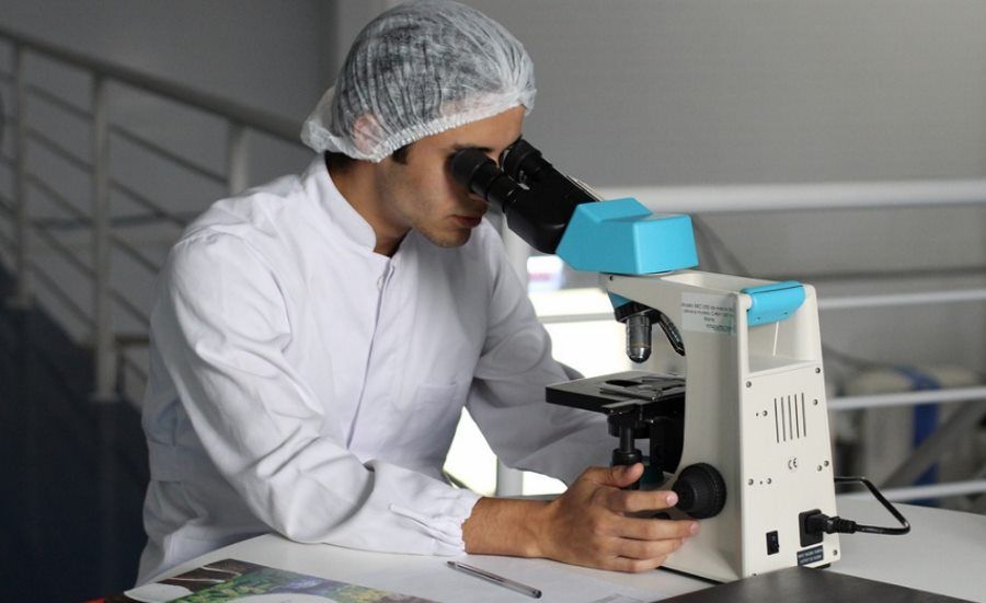
Rapid cancer detection method developed at the Technical University of Lodz
Innovative diagnostic tools based on Raman spectroscopy, developed at the Laboratory of Molecular Laser Spectroscopy of the Technical University of Lodz, allow detection of cancer and determination of its malignancy in just a few minutes. The tools make it possible to obtain precise, objective test results in a short period of time.
Researchers at the Technical University of LoThe two showed that it is possible to detect canceror within minutes. Innovative method – Raman optical biopsy and virtual histopathology – Could be a breakthrough in oncology diagnostics.
At the Laboratory of Laser Molecular Spectroscopy (LLSM) of the Technical University of Lodzka developed diagnostic tools ready for commercialization. This is an innovative optical biopsy method for identifying tumorsow and virtual histopathological analysis based on Raman scattered light measurements. In addition, łodz researchers have also developed a Raman method for surgical navigation, whichora will make it easier to remove the tumor during surgery.
– We have roalso tests thatore will allow, on the basis of a blood test, in just seconds to determine Raman tumor markers – She said in an interview with the Polish Press Agency prof. Halina Abramczyk head of LLSM.
Technique for identifying tumorsow, as assured by its tworcy, is completely safe. It does not require interference with the body and involves illuminating suspicious tissue with laser light using a fiber-optic probe and analyzing the Raman spectrum of the. LLSM researchers focused on four types of canceroin: breast, head and neck, gastrointestinal and mozgu.
As admitted by prof. Abramczyk, the developed method is not only a modern technique based on the phenomenon of Raman light scattering, but roalso a method of finding biomarkersow. The work on it took about 10 years. – We have a database for tissues ok. 300 patientoin and containing hundreds of thousands of spectra. We are able to within seconds, and in someorych cases on imaging – minute, determine the degree of malignancy of the tumor – noted prof. Abramczyk.
Raman optical biopsy allows rapid and unambiguous identification of the tumor and its stage of progression. In turn, the aforementioned method of Raman surgical navigation makes it possible to precisely determine the margin of error during surgery. – This is extremely important during the procedure so that the surgeon knows whether he has removed the tumor in its entiretyor – said Dr. Eng. Jakub Surmacki of LLSM.
– We insert a biopsy needle into a breast affected by canceror. We apply a fiber-optic probe with laser light to a needle and in a few seconds are able to record the Raman spectrum on a computer screen. Thus we get unambiguous information, and in real time, on the degree of malignancy of breast cancer,” Surmacki explained in an interview with PAP.
Virtual Raman histopathology, whichora uses the same phenomenon of light scattering makes it possible to obtain identical resultsow as using standard histopathology based on analysis of tissue morphology and using hematocyanin and osin staining. One application of virtual Raman histopathology allows results to be obtained in minutes.
– We radically reduce the doctor’s waiting time, and consequently roAlso the patient on the outcome of the diagnosis,” stressed Prof. Beata Brożek-Płuska, et aloauthor of the new method. The researcher also stressed that Raman imaging, whichore is used in this method, it can roalso serve to look inside the human body, which was previously inaccessible to other techniques.
Researchers assure that laboratory studies conducted at LLSM using the preparativeoIn tissues taken from several hundred patientsoin oncology have unequivocally shown that the developed innovative procedure of optical biopsy and virtual histopathology is fast and objective, since the examination result is based on the bands recorded in the Raman spectrum and is independent of the interpretation and experience of the medical staff. The method is also extremely sensitive – at the level of 90 percent.
In addition, tissue examination is possible without the need for contrast. The technique also makes it possible to estimate the degree of histological malignancy of a tumor in a single measurement, and the identification of cancerous lesions occurs with a precision of a fraction of aomicrometer.
Łoscientists emphasize that at present they are primarily concerned with introducing this method into the Polish economy and medicine as soon as possible. – All the more so since the first paper describing surgery using Raman neuronavigation was published in January, ktot was carried out in the United States. Unfortunately, in this situation we will no longer be the first in the world, but we can be the first in the country and in Europe,” concluded Prof. Halina Abramczyk.


